

“We are proud of this acquisition, since we have managed to do the first private hospital in Central America in Guatemala to have a comprehensive high precision system for surgical interventions of column, neck and skull. In the pillar we work day by day in the development of technologies that help the care of patients according to modern medicine. The hospital El Pilar is immersed in a digital remodel Vanguard teams that support our doctors from all specialties, ”said Dr. Federico Ranero, medical director of Hospital El Pilar.
New technologies
Advanced O-ARM imaging, allows you to obtain 2D and 3D images in real time, optimizing surgical planning, precise implant placement and postoperative evaluation. The 360 ° images provide a complete vision of the patient. The wide field of vision (40 cm), is ideal for complex procedures and with fluorescence observing microscopic structures that facilitate surgery.
The surgical microscope with intraoperative fluorescence is a micro inspection tool that is used in the Kinevo 900 robotized visualization system. And the qevo tool allows surgeons to inspect areas that cannot be seen directly with a surgical microscope. It is a system that combines advanced optics with digital navigation and high -resolution images that improve surgical accuracy. In addition, it allows in -depth visualization to address the most demanding surgeries.
Among its advantages include:
- Minimal invasion of the column. It does not require large wounds or expose very extensive areas.
- Greater precision when placing materials inside the column, such as boxes and screws.
- It decreases the risk of a reintervention and offers a great contribution in cases of spinal mergers, deformities corrections, spinal decompression and tumor resection.
In addition, spine interventions, this technology can be used in brain diseases, such as tumors, Parkinson’s disease, epilepsy and hydrocephalus. The important thing is adaptability to all types of patients, even those with challenging anatomies, since the clarity of image is obtained, without compromising the surgical workflow.
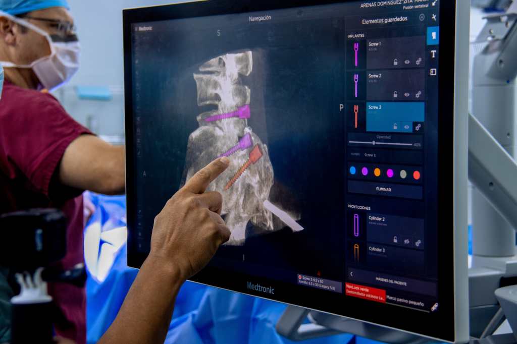
“The neuronaver uses previously made images of magnetic resonance or tomography, which serve as a GPS at the time of performing surgery. When we complement it with the O-ARM we have a 3D image in real time. In this way, we place an implant inside the brain, specifically in the area of the skull, it also helps us to locate the tumors and make a more thorough surgery,” said Dr. Mauricio. Longo, specialist in vascular neurosurgery and skull base surgery.
The incorporation of these devices in column, neck and skull operations contemplate:
- The dynamic visualization in 4K and 3D that allows greater precision.
- Robot -assisted navigation reduces surgeon fatigue and fluorescence technology identifies organs more clearly.
- Better visualization for the surgeon by having a real -time view and safer procedures.
- Potential reduction of blood and pain loss.
- Lower times of stay in the operating room due to preoperative planning.
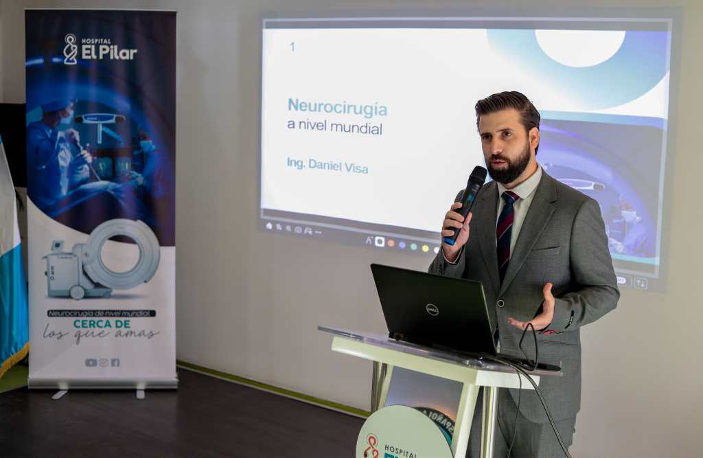
The Pilar has also acquired specialized equipment for advanced neurosurgical procedures. An Allen type surgical table, designed for optimal neurosurgery positioning. The arc in Cios Spin, which offers high quality three -dimensional images during surgery and microcirgery chair, maximizing the ergonomics and comfort of the surgical equipment.
“The objective is to improve the accuracy in surgical interventions and provide visual and quantifiable information for the surgeon, which also becomes multiple benefits for the patient. Accompanied by a multidisciplinary team made up of specialized neurosurgeons, highly trained nursing staff and infrastructure certified internationally,” said Eng. Daniel Visa, executive director of the Hospital El Pilar.